2020 AHA/ACC Guideline for the Diagnosis and Treatment of Patients With Hypertrophic Cardiomyopathy (html) (pdf)
Differential Diagnosis
Left ventricular hypertrophy is commonly seen on echocardiography. Therefore important to know the differential.
Hypertension
Aortic Stenosis
Amyloidosis
Anderson-Fabry Disease
High-level Athelete
High Yield Echo Features
Wall thickness
SAM (Systolic Motion of Mitral Valve)
Mitral Regurgitation
LVOT (Left Ventricular Outflow Track) obstruction
Diastolic Dysfunction
Wall Thickness
Unexplained LV wall of 15mm or more in any wall is pathologic
Commonly hypertrophic cardiomyopathy presents with assymetric. A ratio septal:posterior wall of greater 1.5:1 is pathologic.
Occasionally apex is more effected commonly referred to as apical hypertrophic cariomyopathy
Patient with hypertrophic cardiomyopathy.
Note significant left ventricular hypertrophy.
In the short axis there also is papillary muscle hypertrophy.
SAM (Systolic Anterior Motion of Mitral Valve)
Systolic anterior motion of the mitral valve is commonly seen in patients with HCM.
Since there is a thickened septum blood flow is high and turbulent through the LVOT.
The high flow of blood through the LVOT can draw the mitral valve anteriorly and towards the LV outflow track causing more obstruction. This is known as the Ventruri Effect.
Note the turbulent flow through the LVOT on colour doppler in the parasternal long axis view.
In the zoomed 5 chamber view you can visualize the mitral valve being sucked into the LVOT.
SAM can also be M-Mode through the mitral valve.
Mitral Regurgitation
Commonly seen in Hypertrophic Cardiomyopathy.
A posterior directed mitral regurgitation jet often accompanies SAM.
It temporally follows the onset of LVOT obstruction and care should be taken not to confuse its Doppler velocity profile with that of LVOT obstruction.
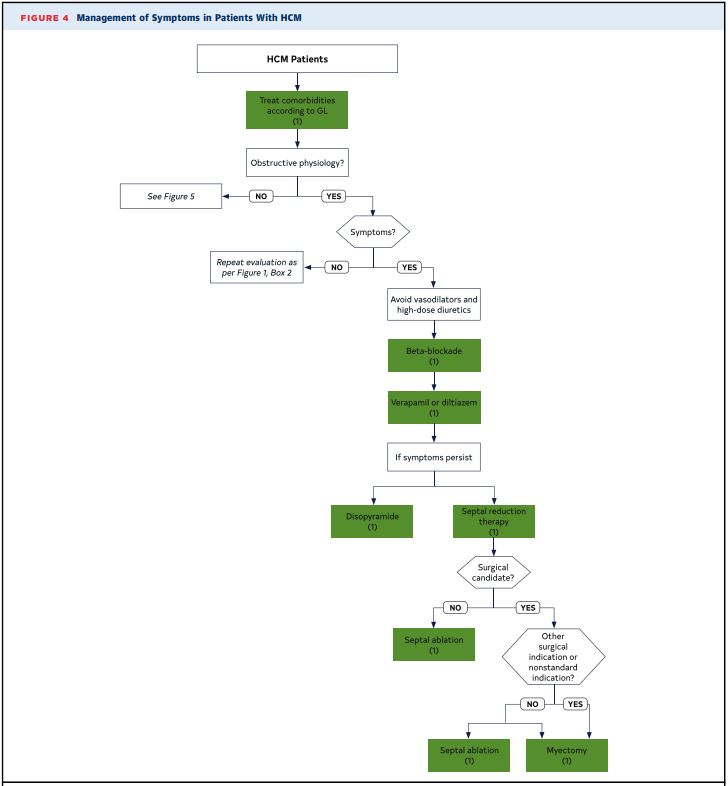
LVOT Obstruction
Pulse wave doppler should be used to localize site of obstruction.
Continuous wave doppler should be used to determine the peak.
If LVOT gradient less than 30 mmHg Valsalva should be attempted.
If no significant LVOT obstruction but patient symptomatic should consider Stress Echo.
Obstructive Physiology defined as a gradient conditions of ≥30 mm Hg.
Marked gradients ≥50 mm Hg, either at rest or with provocation, represent the conventional threshold for surgical or percutaneous intervention if symptoms cannot be controlled with medications.
Pulse wave doppler along the septal wall to localize the site of obstruction.
Note the step-up in gradient.
Commonly HCM PW/CW is described as “dagger shaped”.
Continuous wave doppler through the LVOT demonstrating a peak gradient of 33mmHg at rest
This is considered obstructive physiology.
Diastolic Dysfunction
Diastolic filling of the left ventricle is impaired in about 80% of individuals with HCOM
Can even be observed in asymptomatic patients without overt hypertrophy but who are genetically affected.
No clear relationship exists between the severity of hypertrophy and the severity of diastolic dysfunction.


