Introduction
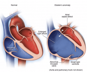
- Apical displacement of the septal and postero-lateral leaflets of the tricuspid valve resulting in:
- “Atrialized” RV above the valve (ratio)
- Small “functional” RV below the valve
- Very large anterior leaflet (attaches the same location while other leaflets shift apically)
- Severe RA enlargement
- Can be cyanotic with an ASD or PFO (frequently associated)
- 5.2 per 100,000 births
- Classic Association: Maternal Lithium use
Associations
- ASD or PFO (50%)
- Accessory Pathway / WPW (25%)
- Pulmonary Stenosis (rare)
Physical Exam
- Inspection: Cool periphery ± cyanosis (poor cardiac output)
- JVP – “V” wave unreliable
- Can have torrential TR and no V-wave (RA is very large and absorbs the regurgitation jet)
- RV Lift Subtle (RV wall is thin)
- Auscultation
- Loud T1 (anterior leaflet large, “flaps”, makes sound)
- Holosystolic murmur worse with inspiration (TR)
- Clicks (large valve clicks when opens)
Diagnosis
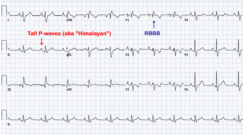
- ECG
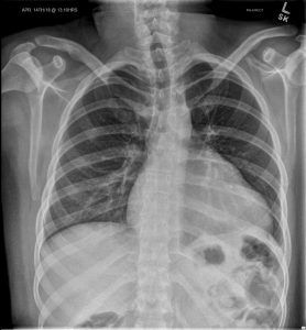
- Chest Xray
- Echo Doppler
- Oximetry
Echocardiogram
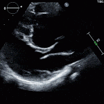
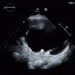
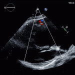
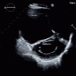
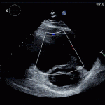
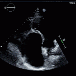
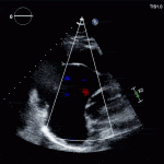
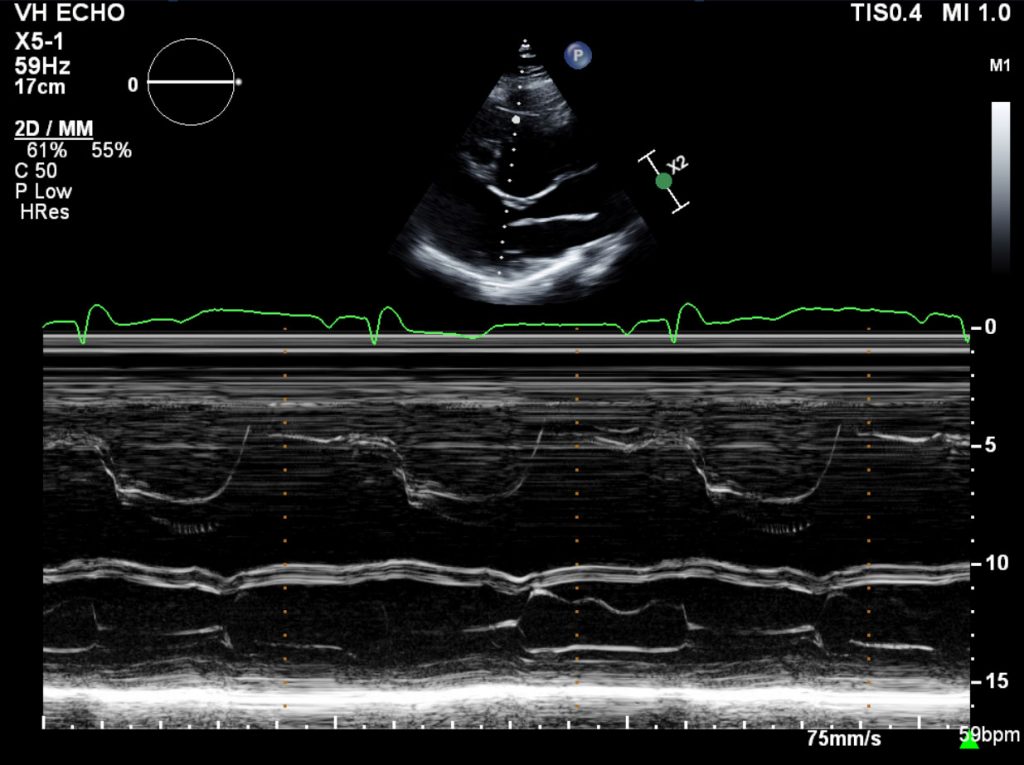
- ***See tricuspid valve in the parasternal long axis***
- Tricuspid and mitral valves are never in the same plane in normal individuals
- Sail-like large redundant anterior leaflet
-
Shiina et al. J Am Coll Cardiol 3(2 Pt 1):356–370, 1984 Diagnostic Criteria Septal leaflet is 8mm/m2 displaced from the crux of the heart (defined by mitral valve insertion)
- RV size and function correlates to disease severity and is prognostic
- The more atrialized the RV, the less functional it is
Management
- Surgical Repair
- Tricuspid valve repair (if feasible) or replacement
- Selective plication of atrialized RV
- Reduction atrioplasty (reducing RA size)
- Arrhythmia Surgery
- Closure of ASD (if present and safe)
- Surgical Issues:
- If RV is too small or severely dilated/dysfunctional such that cannot tolerate full stroke volume, need single-ventricle palliation measures (Glenn/Fontan etc)
- Glenn/Fontan cannot be done if LA pressure or LVEDP are high or LV dysfunction is present (Class IIB – AHA)
- EP study prior to surgery is often done (Class IIB – AHA)
- 1/3 have multiple accessory pathways
- Once valve is replaced, R-sided access for ablation is difficult
- If RV is too small or severely dilated/dysfunctional such that cannot tolerate full stroke volume, need single-ventricle palliation measures (Glenn/Fontan etc)
- Timing of Surgery:
- Children: Offer surgery early if cyanotic (main indication), otherwise wait until older (safer)
- Risk of surgery increases if:
- RV enlarges/fails
- Right Heart Failure / Eisenmenger
- Severe reduction in exercise capacity
| Indications for Repair 2008 (Canadian) |
|---|
| CachNet Guidelines 2008 |
|
| AHA 2018 Guidelines |
| AHA 2018 Guidelines (USA) |
|
Pregnancy
- Pregnancy usually well tolerated
- Contraindications to pregnancy
- Cyanosis
- R-sided HF
- Arrhythmias


