Introduction
- Echocardiography plays a central role in the field of cardio-oncology
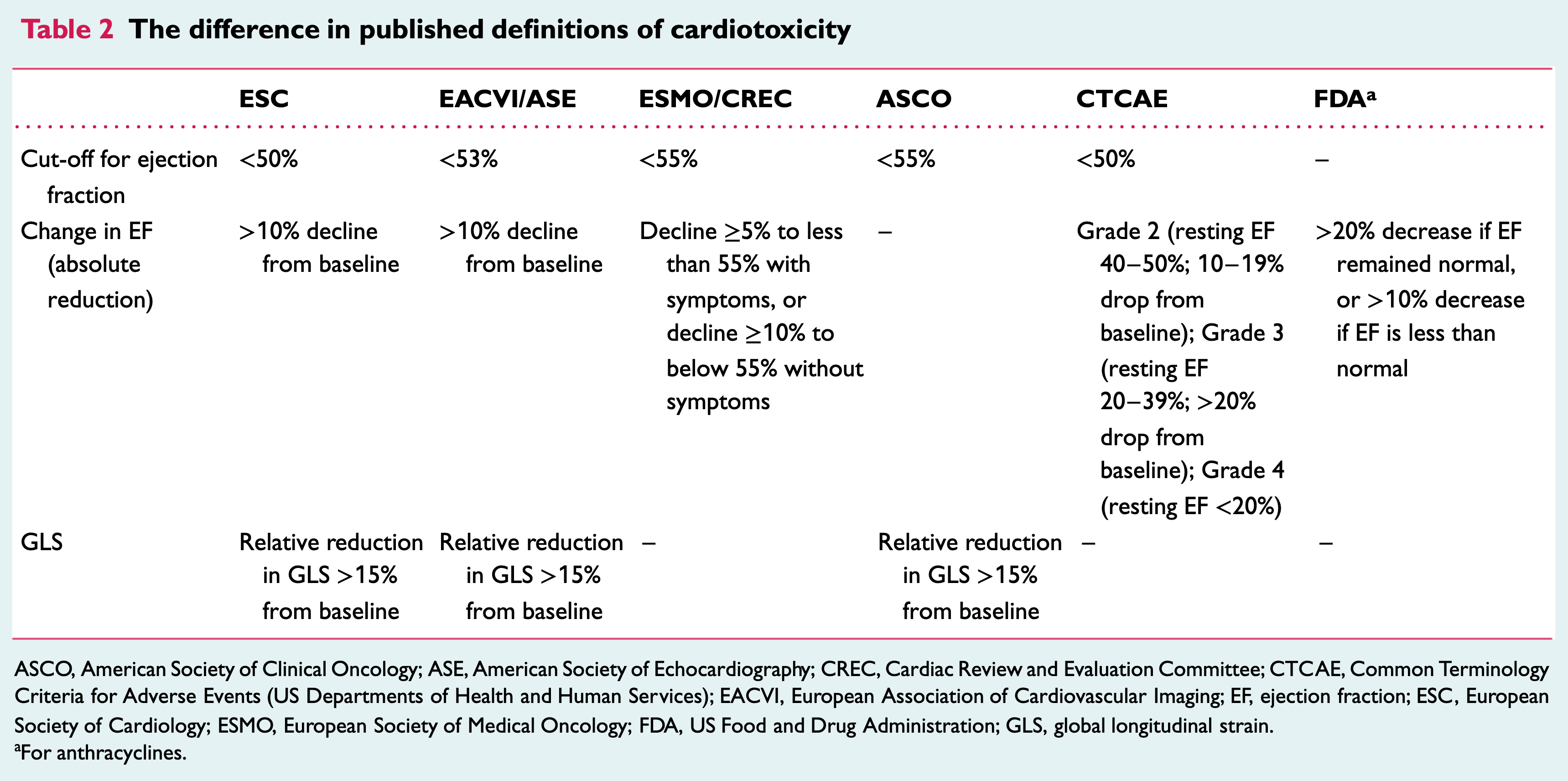
- Many of the trials use LVEF in the definition for cardio-toxicity
- Ongoing area of research to determine how early and how best to predict cardiac complications of cancer therapy
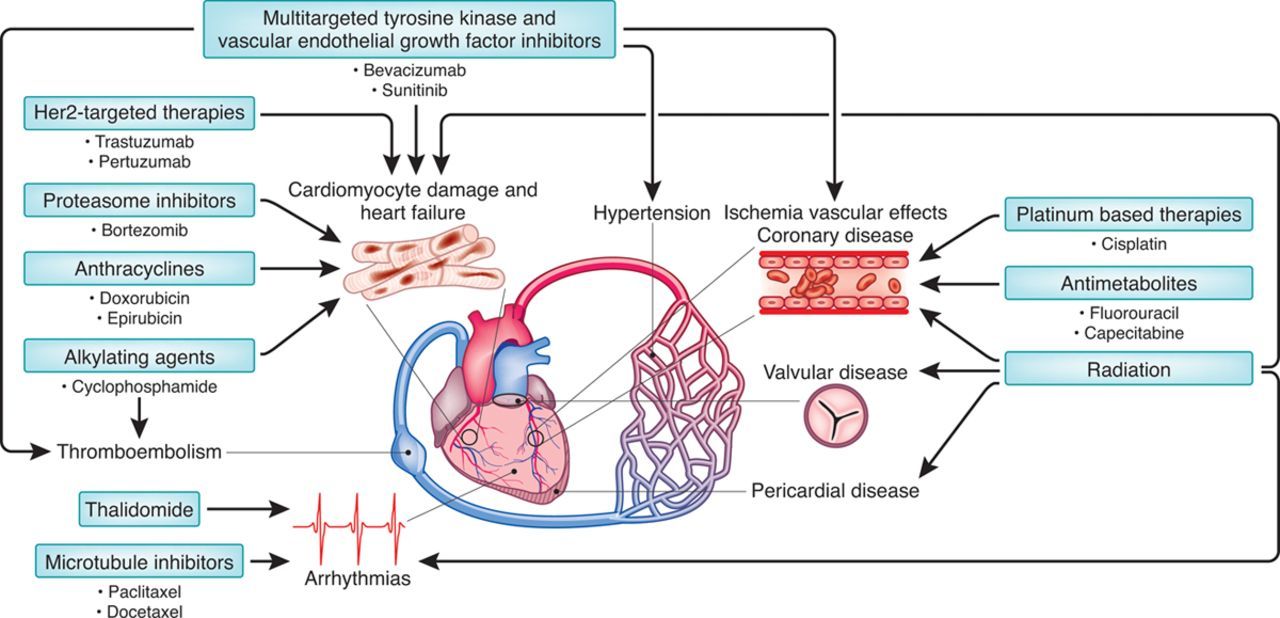
Cardiac Complications of Chemotherapy and Radiation
- Cardiomyopathies
- LV dysfunction (HFpEF or HFrEF)
- Myocarditis
- Takotsubo
- Thrombus
- Infiltration
- Vascular Disease
- Arterial stiffness
- Accelerated athersclerosis
- Pericardial Disease
- Tamponade
- Constriction
- Pulmonary Hypertension
- RV Dysfunction
- Valvular Disease
- Regurgitation
- Stenosis
- Endocarditis
- Arrhythmias
- QTC prolongation
Frequency of Monitoring
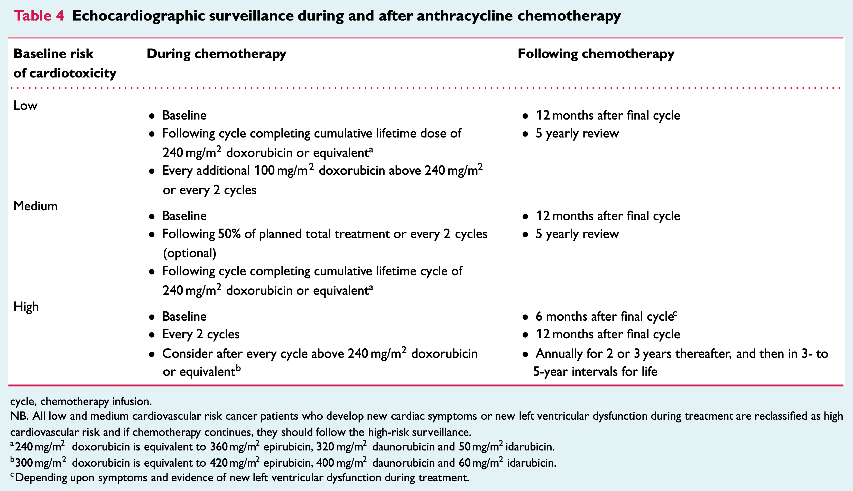
- Surveillance is initially stratified by baseline risk (low, medium, high) and then frequency is determined by chemotherapy cycles while on treatment and monthly after treatment
- Recommendations vary greatly between guidelines
- CCS does not have any recommendation for anthracyclines and recommends q3months for Anti-HER2
- Example surveillance protocols below from ESC
Echo Assessment
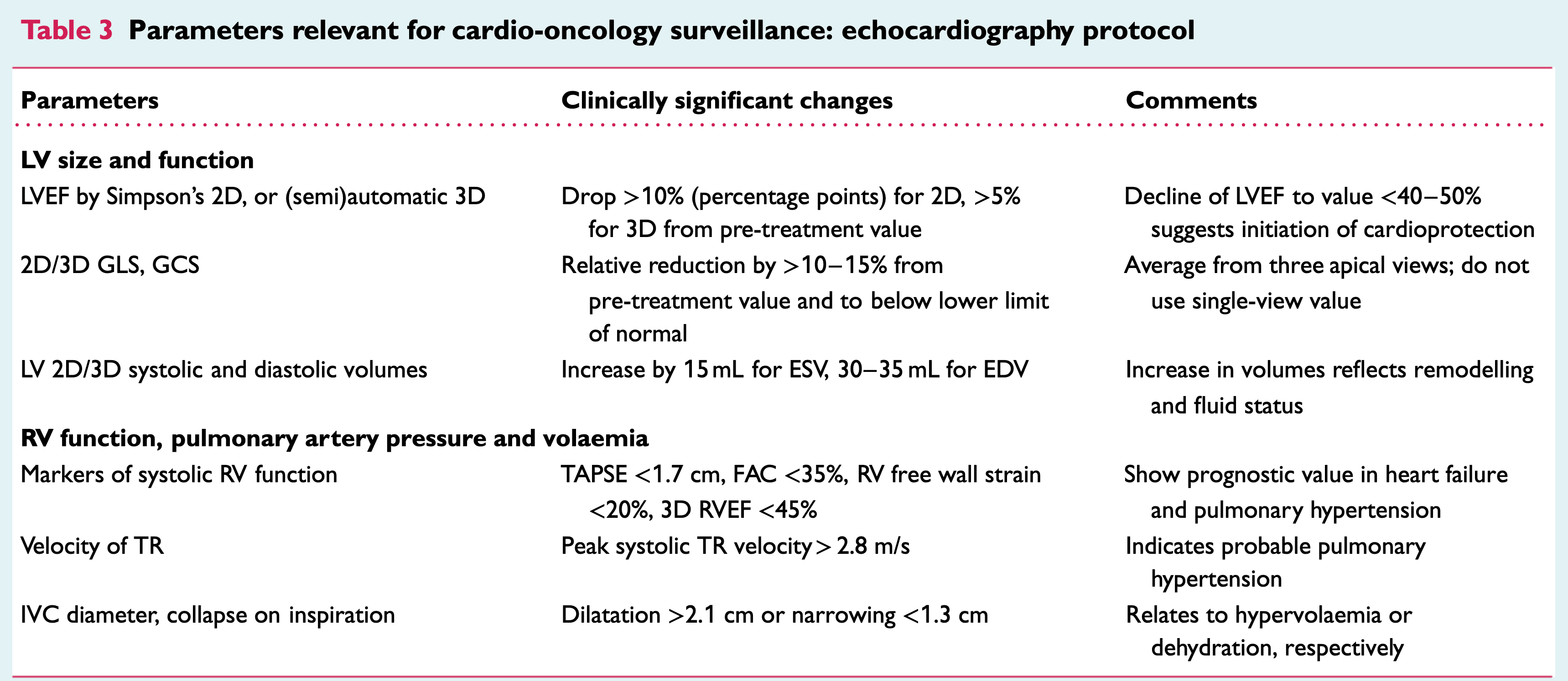
Echo Protocol
LV Systolic Function
2D EF
- Acquisition
- Images should be acquired at end-respiration
- Simpsons method preferred
- In atrial fibrillation use beats with similar R-R interval
3D EF
- Acquisition:
- High-quality ECG with clear R-wave (used for software to trigger full volume)
- Recommend breath hold with shallow breathing, preferably end-expiration (if deep inspiration needed, recommended to document and recreate at next exam)
- Review images before patient patient leaves to look for stitch artifact and ensure if contouring possible
- Some variations between software thus should use same machine and analysis software for serial echo
- Sensitive to image quality thus 2D and 3D images should be taken
- Benefits:
- No assumptions made about LV geometry (ie. short axis view of ventricle is circular)
- Ability to discriminate smaller % changes (5-8%)
- Semi-automated and thus better intra- and interobserver variability
Contrast
- Poor endocardial definition can occur in patients post mastectomy, chest radiation or breast reconstruction surgery)
- Contrast should be used when 2 contiguous LV segments from any apical view are no adequately visualized
LV Diastolic Function
- Diastolic dysfunction may appear earlier than systolic dysfunction so important to acquire on each exam
Global Longitudinal Strain
- Acquisition:
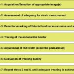
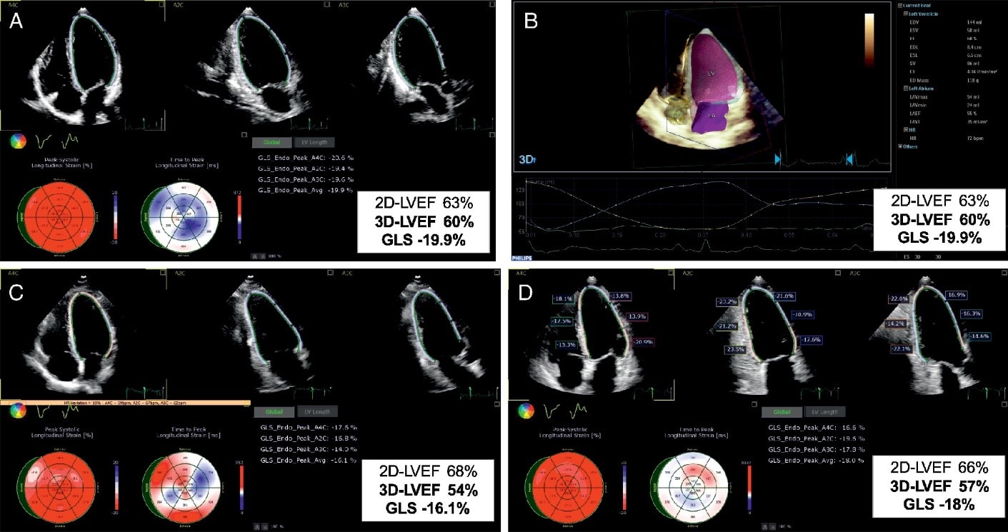
- GLS is calculated using A3C/A4c/A2c views with optimal image quality and frame rate 40-90
- If more than 2 segments have poor tracking, GLS should not be calculated
- Some variations between software thus should use same machine and analysis software for serial echo
- Great article to learn how to acquire strain: JACC 2015: Practical Guidance in Echocardiographic Assessment of Global Longitudinal Strain
- Interpretation
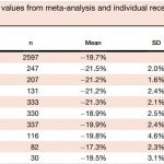
- ASE 2015: “peak GLS in the range of -20% can be expected in a healthy person, and the lower the absolute value of strain is below this value, the more likely it is to be abnormal”
- Cardiotoxicity defined as relative reduction in GLS > 15% from baseline
- Currently being studied how well GLS can predict cardiotoxicity (SUCCOUR study + commentary)
Right Heart
- Not officially included in the definition of cancer treatment related cardiac dysfunction
- Recommended parameters as expected: RV dimenions, RV S’, TAPSE, TR Velocity, FAC
- Certain cancer treatments can cause PH (dasatinib) and/or RV dysfunction (anthracycline, anti-HER2, cyclophosphamide, dasatinib)
Radiation Related Cardiac Dysfunction
- Pericardial disease is the most frequent radiation related complication
- Can occur months to years later
- Should assess for constriction and pericardial effusion
- Valvular disease can occur with radiation
- Fibrotic process which can cause leaflet thickening, shortening and calcifications that may result in stenosis or regurgitation
- More often left-sided valves
- Recommended to image 5 years after RT in high risk patients and 10 years in non high risk followed by q5years
Multimodal Imaging
- CT
- Useful in non-invasive evaluation of coronary disease, pericardial disease, and cardiac tumours
- MRI
- Useful in patients with poor echocardiographic windows
- Great inter- intrareader variability and thus sensitive in detecting small changes in EF
Tutorials
Phillips EPIQ Strain Tutorial
Phillips Dynamic Heart Model (Single-Beat) Tutorial
Case
Further Reading
- 2020 HFA/EACVI/ESC: Role of cardiovascular imaging in cancer patients receiving cardiotoxic therapies (html) (pdf)
- 2021 BSE/BCOS: Guideline for Transthoracic Echocardiographic Assessment of Adult Cancer Patients Receiving Anthracyclines and/or Trastuzumab (html) (pdf)
- 2015 ASE/EACVI: Recommendations for Cardiac Chamber Quantification by Echocardiography in Adults (html) (pdf)
- 2015 JACC: Practical Guidance in Echocardiographic Assessment of Global Longitudinal Strain (html) (pdf)
Authors
- Author: Atul Jaidka (MD, FRCPC, Cardiology Fellow)
- Last Updated: March 24, 2021
- Comments or questions please email feedback@cardioguide.ca
