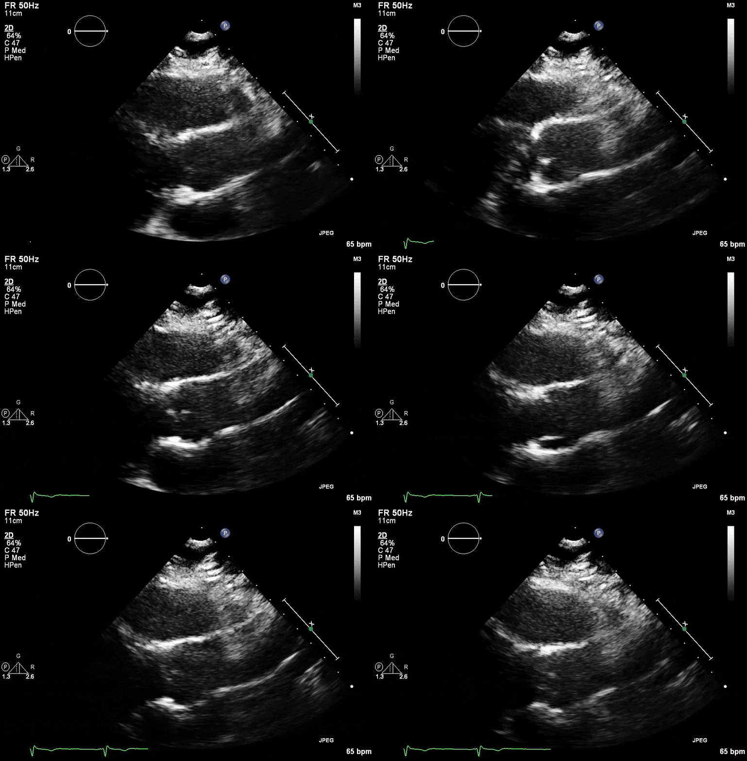
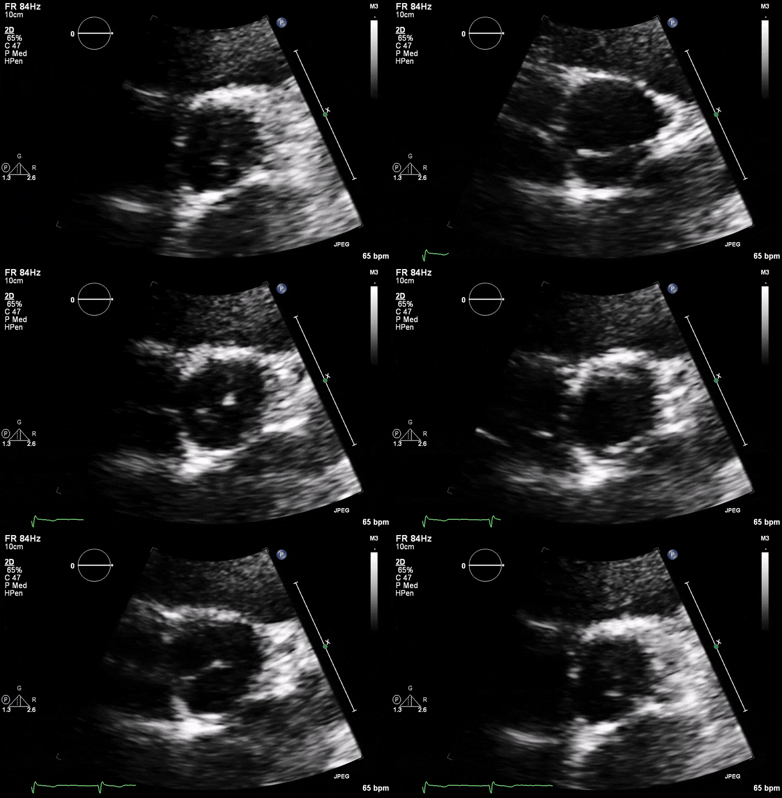
- Most common congenital heart disease abnormality involving 1-2% of the population.
- Strong risk factor for developing early aortic stenosis, aortic regurgitation and is associated with aortopathy such as aortic dissection and aneurysm.
- Should screen all first degree relatives.
Types
- Fusion of right and left leaflet most common usually accounts for 80% of cases.
- Right and non-coronary leaflet the next most common usually 20% of cases.
- Fusion of Left with non-coronary is very rare.
- Unicuspid valve is also very rare.
Role of Echo
- Crucial for diagnosis and characterizing the leaflets involves.
- Important for identifying associated diseases such as aortic aneurysms, associated aortic stenosis or regurgitation.
- Bicuspid valves are best visualized in the short axis view as this allows for the best characterization of the involved leaflets. Long axis views may give clues to the presence of a bicuspid valve as the valve closure line may be eccentric.


