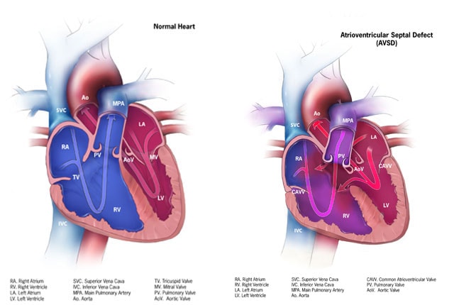
Complete AV Septal Defect Introduction Partial AV Canal Defect –> aka Primum ASDComplete AV Canal Defect ASD above the valveVSD below the valveInstead of 2 valves –> One large AV valve Comparison of normal and complete AVSD anatomy. (CDC Public Domain) Diagnosis Parasternal Long Parasternal Long Color Parasternal Short Axis Parasternal Short Axis Color Apical 4 Chamber View Apical 4 Chamber Color Apical 3 Chamber Subcostal Complicating Features Cleft AV ValveLVOT Obstruction (“Gooseneck Deformity”)Aortic valve positioned anteriorly and farther from the apex than the mitral valve (normally equidistant from apex).Long LVOT can obstruct, this is known as the “Gooseneck Deformity”AV Block Management Usually associated with a massive shunt that needs repair in early childhood.Without intervention, the large shut causes increase in pulmonary flow, which raises pulmonary vascular resistance causing Eisenmenger syndrome.A PA-band may be used to protect the pulmonary circulation from rising vascular resistance until definitive repairSurgical RepairComplete repair can be attempted to separate the AV valves, atria and ventriclesIf RV is not well developed enough –> Single ventricle palliation will need to be performed (Bidirectional Glenn –> Fontan) Case #2 Parasternal Long-Axis Apical 4 Chamber View Apical 4 Chamber AV Color Parasternal Short Axis – AV Valve Post author:Pavel Antiperovitch Post published:November 19, 2019 Post category:Echo / Echo Topics / Pericardial, Myocardial, Pulmonary, Congenital Heart Disease